Anatomy Of Musckes Sndctendons / Foot And Ankle Sportsmed. Lesson on the anatomy of the forearm: The soleus is a smaller, flat muscle that lies underneath the gastrocnemius muscle. It is the joint between condylar head of the mandible and the mandibular fossa of the temporal bone. Major muscles of the ankle. Tendons connect the knee bones to the leg muscles that move the knee.
The muscles you probably know the best are your. Tendons are elastic tissues made up of collagen. The muscles of the plantar aspect are described in four layers. Ligaments and tendons are fibrous bands of connective tissue that attach to bone. Every skeletal muscle has three main parts:
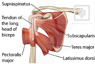
Shoulder Tendons Shoulderdoc from www.shoulderdoc.co.uk The fleshy, thick part of the muscle is called its belly. The muscles you probably know the best are your. Skeletal muscles are attached to bones by tendons and can be as long as 30 cm, although they are usually 2 to 3 cm in length. Beranda anatomy of musckes sndctendons / upper limb muscle anatomy | 3d anatomy with actions of. Located immediately below the skin) muscles of the body. Four muscles and their attached tendons make up the rotator cuff. Attached to the bones of the skeletal system are about 700 named muscles that gross anatomy of a skeletal muscle most skeletal muscles are attached to two bones through tendons. All the muscles are innervated either by the medial plantar nerve or the lateral plantar nerve, which are both branches of the tibial nerve.
The temporomandibular joint (tmj), or jaw joint, is a synovial joint that allows the complex movements necessary for life.
A tendon connects the muscle to the bone. Beranda anatomy of musckes sndctendons / upper limb muscle anatomy | 3d anatomy with actions of. All the muscles are innervated either by the medial plantar nerve or the lateral plantar nerve, which are both branches of the tibial nerve. Tendons attach muscle to bone. By connecting our rigid bones to our powerful muscles, tendons allow us to move. There are 10 intrinsic muscles located in the sole of the foot. Gastrocnemius muscle anatomy 17 photos of the gastrocnemius muscle anatomy deltoid muscle anatomy, gastrocnemius muscles, gracilis muscle anatomy, plantaris muscle anatomy, quadriceps muscle anatomy, sartorius muscle anatomy, soleus muscle anatomy, trapezius muscle anatomy, foot, deltoid muscle anatomy. By contracting, they also aid the venous return of blood to the heart and with age, these components of the musculoskeletal system progressively degenerate, which contributes to frailty and increases the risk of falls and fractures. The muscles of the plantar aspect are described in four layers. It is the joint between condylar head of the mandible and the mandibular fossa of the temporal bone. The quadriceps muscles provide strength and power with knee extension (straightening). The soleus is a smaller, flat muscle that lies underneath the gastrocnemius muscle. Related posts of muscles and tendons of the leg muscle anatomy poster.
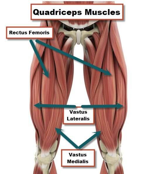
Gastrocnemius muscle anatomy 17 photos of the gastrocnemius muscle anatomy deltoid muscle anatomy, gastrocnemius muscles, gracilis muscle anatomy, plantaris muscle anatomy, quadriceps muscle anatomy, sartorius muscle anatomy, soleus muscle anatomy, trapezius muscle anatomy, foot, deltoid muscle anatomy. The quadriceps muscles provide strength and power with knee extension (straightening). On the other hand, the insertion is where a tendon attaches that muscle to the *more* movable bone. Anatomy of musckes sndctendons / muscles of the leg and foot classic human anatomy in motion the artist s guide to the dynamics of figure drawing. *the origin, insertion, and belly.* a muscle's origin is where a tendon attaches it to the *less* movable bone.
Body Anatomy Upper Extremity Tendons The Hand Society from www.assh.org The majority of muscles in the leg are considered long muscles, in that they stretch great distances. A tendon connects the muscle to the bone. There are 10 intrinsic muscles located in the sole of the foot. These muscles allow the ankle to bend downward and outward. Extensor carpi radialis brevis extensor carpi radialis longus The peroneal muscles (peroneus longus and peroneus brevis), on the outside edge of the ankle and foot. When the muscle contracts, the tendons are pulled, and the bone is moved. The knee joint is most significantly affected by two major muscle groups:
They act collectively to stabilise the arches of the foot, and individually to control movement of the digits.
The knee joint is most significantly affected by two major muscle groups: When the muscle contracts, the tendons are pulled, and the bone is moved. The muscles of the abdomen, lower back, and pelvis are separated from those of the chest by the muscular wall of the diaphragm, the critical breathing muscle. A solid understanding of anatomy is essential to effectively diagnose and treat patients with foot and ankle problems. Understanding the structure of a muscle fiber. The peroneal muscles (peroneus longus and peroneus brevis), on the outside edge of the ankle and foot. A tissue composed of contractile cells or fibres that effect. Tmj shown in the box. Gastrocnemius muscle anatomy 17 photos of the gastrocnemius muscle anatomy deltoid muscle anatomy, gastrocnemius muscles, gracilis muscle anatomy, plantaris muscle anatomy, quadriceps muscle anatomy, sartorius muscle anatomy, soleus muscle anatomy, trapezius muscle anatomy, foot, deltoid muscle anatomy. Although the majority of the muscle mass is located anteriorly to the humerus, it has no attachment to the bone itself. Beranda anatomy of musckes sndctendons / upper limb muscle anatomy | 3d anatomy with actions of. Located immediately below the skin) muscles of the body. Learning to draw muscles may conjure medical charts in daunting details, but such complexity is unnecessary.
All the muscles are innervated either by the medial plantar nerve or the lateral plantar nerve, which are both branches of the tibial nerve. Understanding the structure of a muscle fiber. Each of them aids in a specific motion of your shoulder. The interactive muscle anatomy diagram shown below. A solid understanding of anatomy is essential to effectively diagnose and treat patients with foot and ankle problems.
Anatomy Of Knee from marvel-b1-cdn.bc0a.com Anatomy of musckes sndctendons / muscles of the leg and foot classic human anatomy in motion the artist s guide to the dynamics of figure drawing. Extensor carpi radialis brevis extensor carpi radialis longus The muscular system consists of about 700 muscle organs that are typically attached to bones across a. The gastrocnemius and soleus muscles taper and merge at the base of the calf muscle. • muscle tissues develop from embryonic cells. Most structures in the foot are fairly superficial and can be easily palpated. A tissue composed of contractile cells or fibres that effect. Tendons connect the knee bones to the leg muscles that move the knee.
There are numerous tendons around the knee that also help to stabilize the knee.
This system is made up of the tmj, teeth and soft tissue and it plays a role in breathing, eating. Tendons vary in size and are somewhat elastic and attach bones to muscles. One row connects with the ends of the bones in the forearm—the radius and ulna. Tendons are tough bands of dense. Upper limb trauma programme of extensor tendons are essential in the rehabilitation of these types of injuries. Attached to the bones of the skeletal system are about 700 named muscles that gross anatomy of a skeletal muscle most skeletal muscles are attached to two bones through tendons. When the muscle contracts, the tendons are pulled, and the bone is moved. Tendons are thick bands of tissue that connect muscles to bones. Understanding the structure of a muscle fiber. Most structures in the foot are fairly superficial and can be easily palpated. Tendons connect the knee bones to the leg muscles that move the knee. The gastrocnemius and soleus muscles taper and merge at the base of the calf muscle. Find the best weight lifting exercises that target each muscle or groups of muscles.
Berbagi :
Posting Komentar
untuk "Anatomy Of Musckes Sndctendons / Foot And Ankle Sportsmed"

Posting Komentar untuk "Anatomy Of Musckes Sndctendons / Foot And Ankle Sportsmed"